CT scan for a single cell
Scientists create the first 3D images of a living
cell.
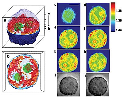
C thru J are 2D scan images used to produce 3D A & B
A new imaging technique developed at MIT has allowed scientists to create the first 3D images of a living cell, using a method similar to the X-ray CT scans doctors use to see inside the body.
The technique could be used to produce the most detailed images yet of what goes on inside a living cell without the help of fluorescent markers or other externally added contrast agents.
The current resolution of the new technique is about 500 nanometers, or billionths of a meter, but the team is working on improving the resolution. “We are confident that we can attain 150 nanometers, and perhaps higher resolution is possible,” Michael Feld said.
I wonder if it can see how many pinheads are dancing on an angel?
Posted: Thu - August 16, 2007 at 08:57 AM