Molecular structure of influenza
Scientists…have succeeded in imaging, in
unprecedented detail, the virus that causes influenza.
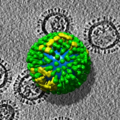
Virus is about 120 nanometers
Scientists…have succeeded in imaging, in unprecedented detail, the virus that causes influenza.
A team of researchers led by Alasdair Steven, Ph.D., working with a version of the seasonal H3N2 strain of influenza A virus, has been able to distinguish five different kinds of influenza virus particles in the same isolate (sample) and map the distribution of molecules in each of them. This breakthrough has the potential to identify particular features of highly virulent strains, and to provide insight into how antibodies inactivate the virus, and how viruses recognize susceptible cells and enter them in the act of infection.
The research team used electron tomography (ET) to make its discovery. ET is a novel, three-dimensional imaging method based on the same principle as the well-known clinical imaging technique called computerized axial tomography, but it is performed in an electron microscope on a microminiaturized scale.
I spent a couple of very happy years working in photo-micrography — long before this sort of resolution was possible. Amazing stuff.
Posted: Mon - January 8, 2007 at 06:24 AM