New Detailed Images of Virus
Fifty years after MIT researchers pioneered the
use of electron microscopy to study viruses, MIT scientists have helped produce
the most detailed images yet of the tiny infectious agents.
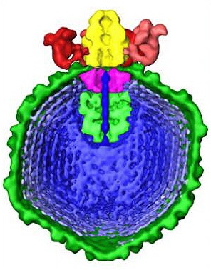
The big blue critter with green skin — is the virus.
Fifty years after MIT researchers pioneered the use of electron microscopy to study viruses, MIT scientists have helped produce the most detailed images yet of the tiny infectious agents.
The images, which show for the first time a virus poised to inject its genetic material into a host cell, grace the cover of the Feb. 2 issue of Nature.
Scientists have known for decades that viruses infect cells by injecting their genetic material, either DNA or RNA, into host cells, but even with electron microscopy, “we could never see the details of that aspect of it,” said Jonathan King, an MIT professor of biology and one of the authors of the paper.
This project builds on a long legacy of viral research at MIT, King said. In 1969, MIT Professor Salvador Luria shared the Nobel Prize in physiology or medicine with Max Delbruck and Alfred Hershey for work on the genetic structure and replication mechanisms of viruses.
Bravo!
Posted: Thu - February 16, 2006 at 07:31 AM